↗ PT

Other pages are available in Portuguese

Skip to content

Cavities, stains and cracks

A stain, a line in the enamel or pain when chewing may have different causes. Before whitening, restoring or treating the inside of the tooth, its origin, depth and activity need to be understood. [1] [3] [4]

Book an assessment

The same tooth. Three assessments.

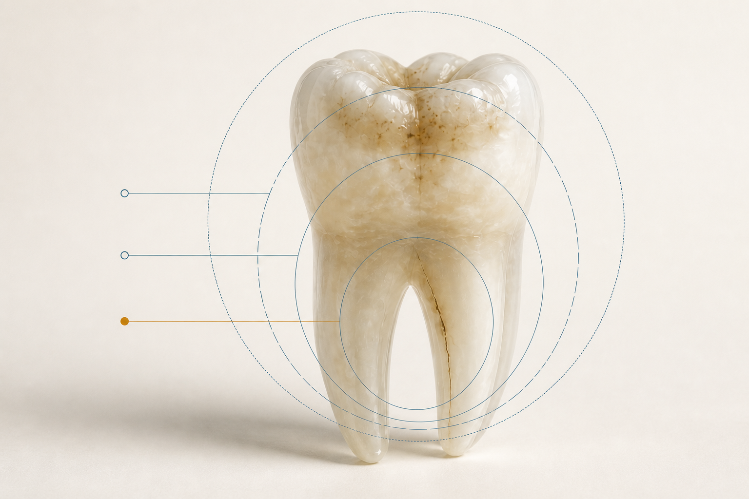
Conceptual artwork of a tooth seen through three observation lenses. It does not represent a patient, examination or diagnosis.

Colour, texture, sensitivity and history need to be considered together. No isolated detail determines the approach.

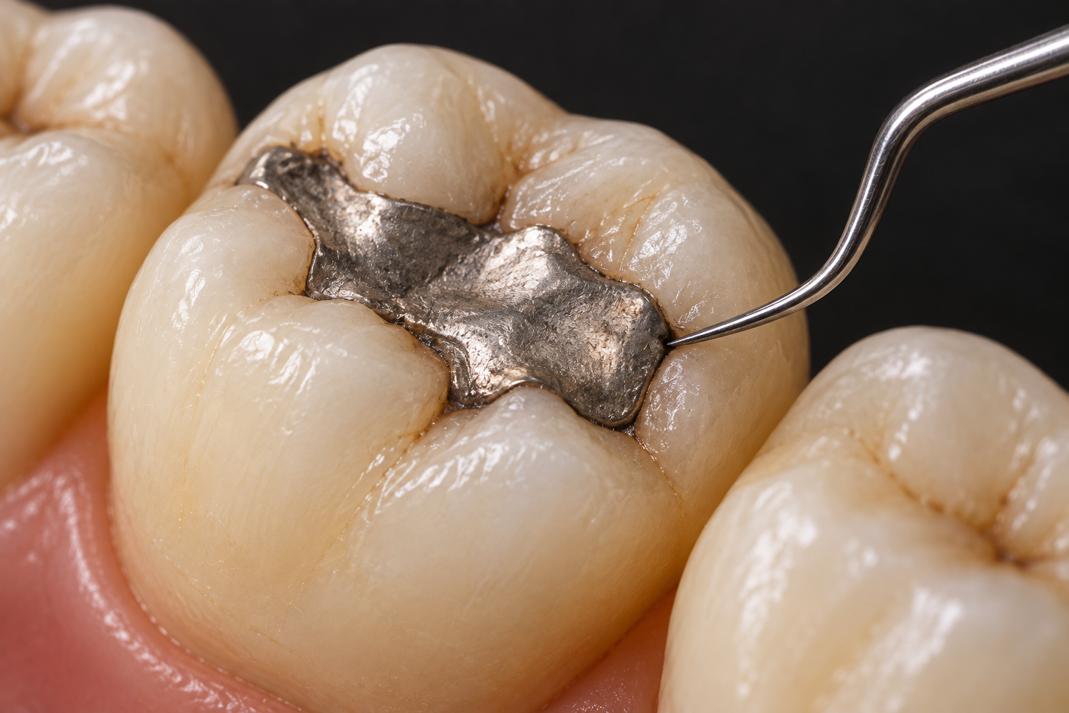
Decay

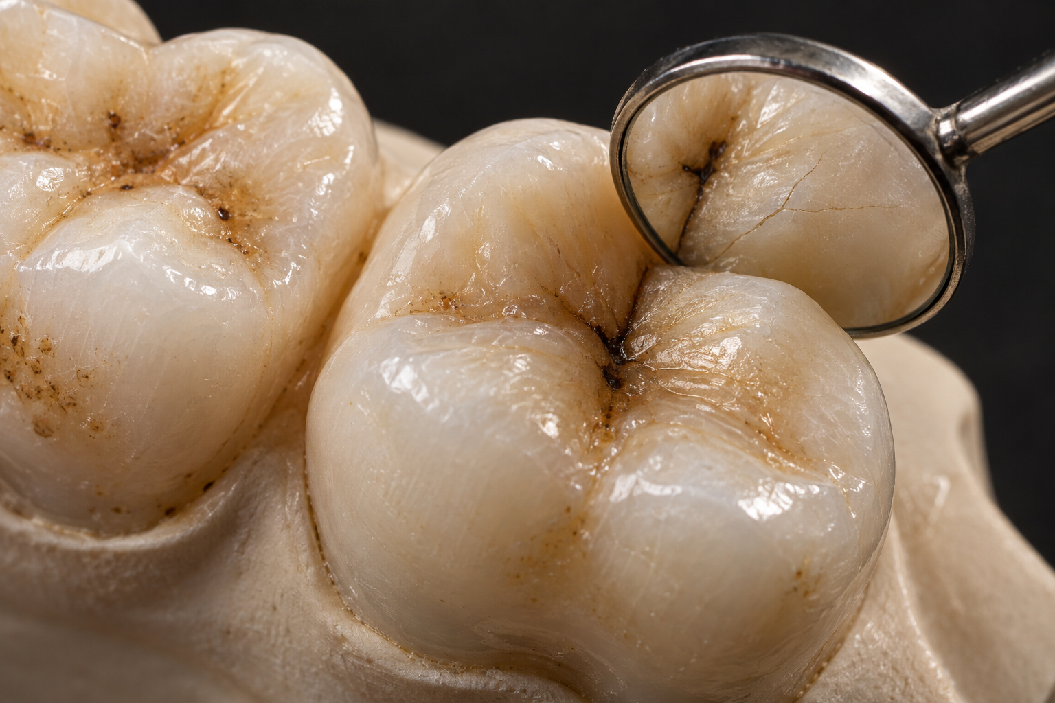
It does not simply mean a hole. A lesion may be at an early stage without cavitation or may already involve loss of structure. Activity, depth, surface and individual risk change the decision. [1] [2]

Stain

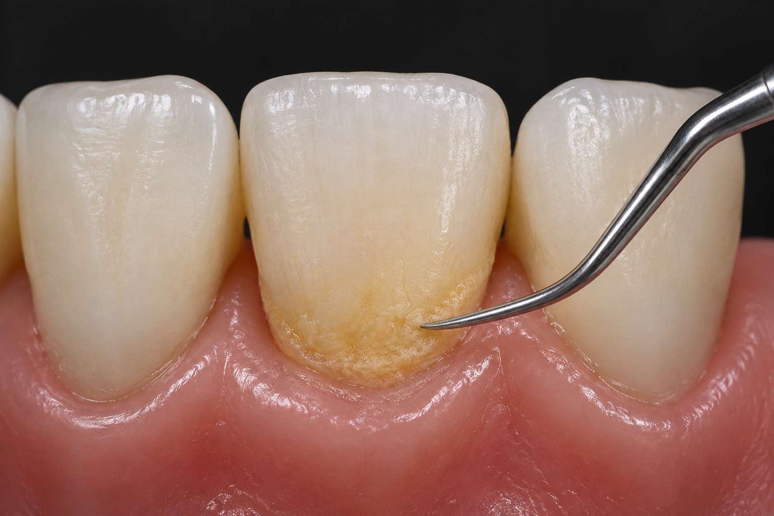
Pigments may remain on the surface, originate inside the tooth or enter through enamel changes. The cause affects what can be cleaned, whitened, monitored or restored. [4]

Crack

Superficial lines, fractured cusps and deep cracks do not have the same significance. Type, location, extent and symptoms guide prognosis and treatment. [3]

Assessment changes the focus.

Surface
Colour, shine, texture, plaque, restorations and visible loss of structure.
Response
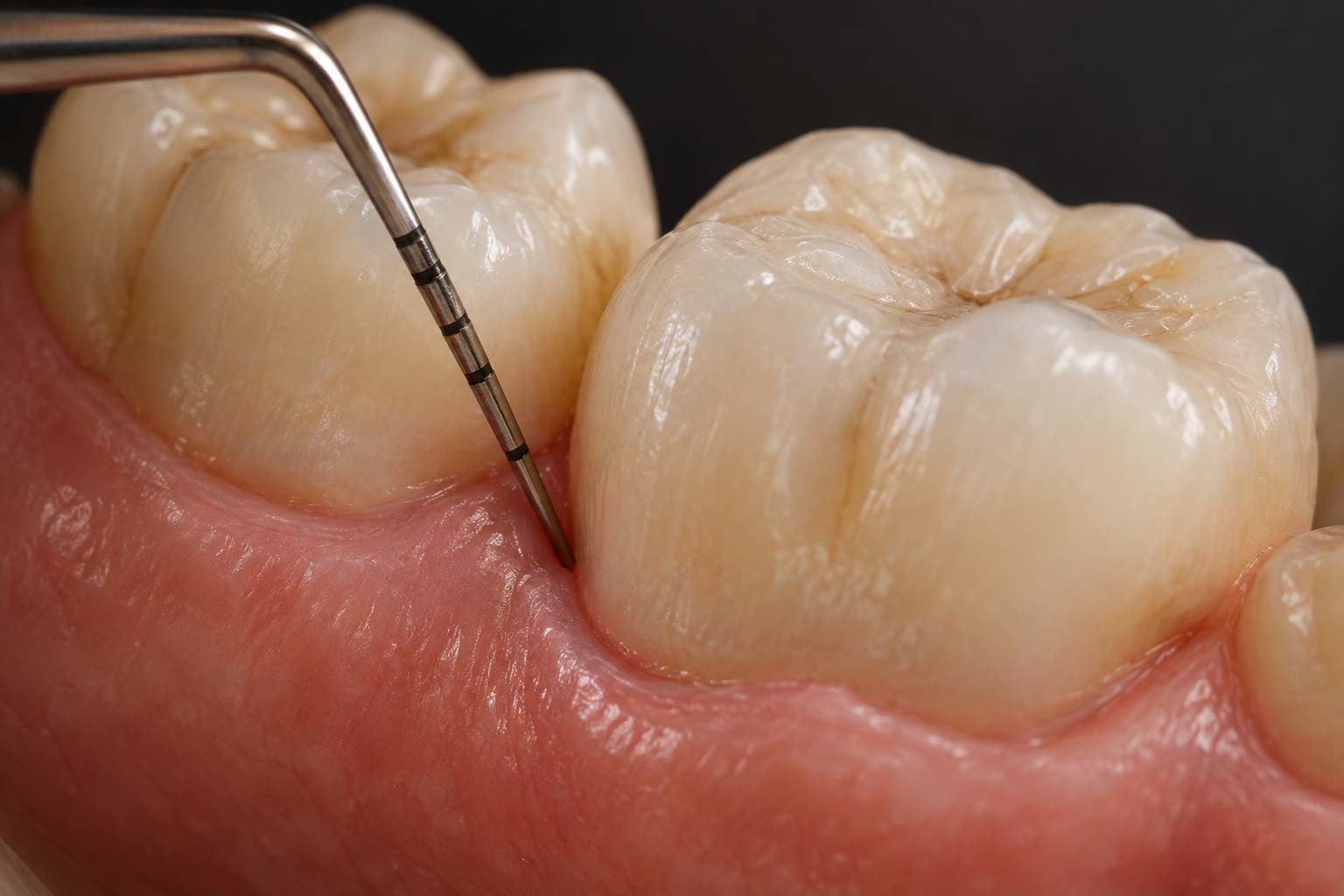
Sensitivity to cold, heat, sweetness, pressure and releasing the bite.
Context
When it began, whether it has changed, whether there was trauma and which habits or treatments are already present.
Additional information
Tests and imaging are selected when they add information. No examination replaces the full clinical assessment.

Preserving the tooth guides the choice.

When structure allows, the plan seeks to control the cause and preserve tooth tissue. This does not mean delaying care. It means choosing an intervention consistent with the diagnosis. [1] [2]

  1. Follow up

    Some findings without activity or clinical consequences may be recorded and monitored.

  2. Control

    Decay lesions may receive non-restorative measures in specific situations, with reassessment.

  3. Protect

    A weakened structure may need protection to reduce loading and limit progression.

  4. Restore

    When structure has been lost, restoration aims to restore shape, sealing and function while preserving what is possible.

  5. Treat the inside

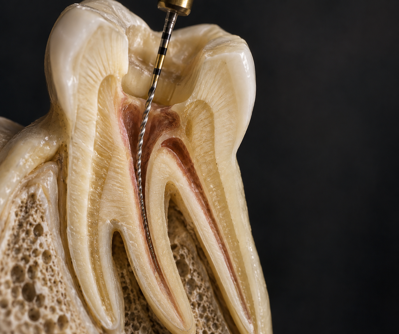
    If the pulp is involved, endodontic assessment may form part of the plan to try to retain the tooth.

Some signs need attention without waiting.

Severe or spontaneous pain, swelling, recent trauma, loss of part of a tooth, pain on biting or intense, persistent sensitivity warrant assessment. In cracked teeth, pain may come and go, and its origin is not always straightforward to locate. [3]

  • Pain on pressure or on releasing the bite
  • A sharp response to cold or heat that does not quickly pass
  • A broken piece or sharp edge
  • Swelling, discharge or spontaneous pain

Primary and permanent teeth need their own decisions.

In children, age, cooperation, the expected time that tooth will remain in the mouth and how soon the primary tooth is expected to fall out naturally form part of the plan. In permanent teeth, lesion extent, vitality, remaining structure and the possibility of follow-up also matter.

Decay guidelines distinguish primary from permanent teeth and consider specific clinical scenarios. [1] [2]

Before choosing treatment.

Direct answers to common questions. Actual suitability depends on individual examination.

Is every dark stain decay?

No. Surface pigmentation, internal changes, old restorations and decay may change colour. Appearance needs to be related to texture, activity, depth and context. [4]

Could a white spot be decay?

It may represent an early mineral change, but other enamel conditions also create white areas. Assessment distinguishes origin and activity before recommending an approach.

Does all decay need a restoration?

No. Depending on stage and surface, non-restorative approaches can control or arrest lesions. Cavitation, depth and individual risk change the decision. [1] [2]

Does a cracked tooth always hurt?

No. Some lines are limited to enamel and cause no pain. Others may cause pain when chewing or thermal sensitivity. Type, location and extent determine their significance. [3]

Does a crack close by itself?

Not like a bone. Treatment seeks to protect the tooth, control symptoms and reduce the chance of progression, but prognosis varies and no protection guarantees absolute success. [3]

Can I whiten before investigating a stain?

It is best to understand the cause and check the health of teeth and restorations first. Pigments from different origins do not respond in the same way.

Is treatment the same in children and adults?

Not necessarily. Tooth type, developmental stage, decay risk, structure and the possibility of follow-up influence the choices.

How long does it take?

There is no single timeframe. Cleaning surface pigment, controlling a decay lesion and caring for a deep crack are different situations. Timing only makes sense after diagnosis.

What happens if tooth decay is not treated?

Decay can advance from the enamel to the inner layers of the tooth. Over time, it can cause pain, reach the pulp, lead to infection and result in tooth loss. The speed varies from person to person, and there is not always pain at the start. That is why regular check-ups matter. [5]

What causes tooth decay?

Some plaque bacteria use the sugar in food to produce acids, which remove minerals from the tooth. Frequent sugar intake, insufficient hygiene, little saliva and little contact with fluoride increase the risk. The assessment considers these factors for each person. [5]

Can tooth decay be cured?

At the start, before a cavity forms, decay can be stopped and even reversed with fluoride, saliva and control of plaque and sugar. When structure has already been lost, the tooth does not rebuild itself, and treatment aims to control the lesion and restore what was lost. [2] [1] [5]

How much does treating decay, stains or cracks cost?

The cost depends on the diagnosis, the number of teeth, the material and the type of care indicated. It is presented after the assessment. Treatment can be paid in up to 21 credit card instalments, depending on the treatment plan and current conditions.
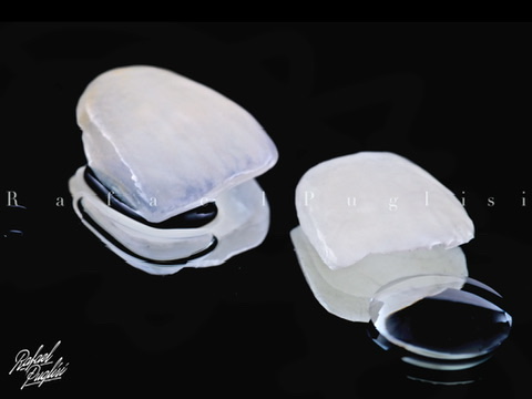
Beyond aesthetics, we are a multidisciplinary clinic.

Explore options

Care takes place here

Across more than 400 m², seven consulting rooms and an integrated in-house laboratory bring together different stages of care at the same address. Discover the Institute.

A history in dentistry since 1923 connects generations with care today. Explore the Institute's history.

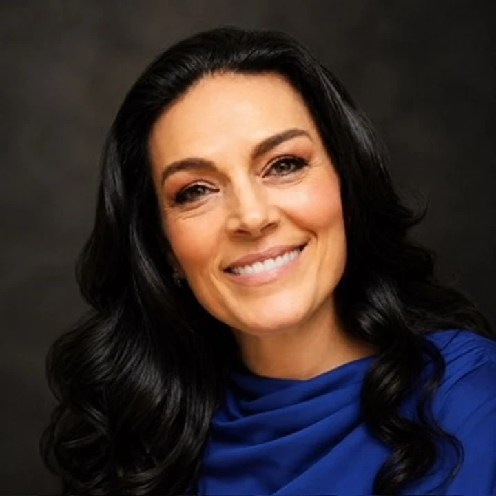
Documentary portrait of Dr Silvana Puglisi

Dr Silvana Puglisi is the dentist responsible for technical oversight. Meet our professionals who continue to provide care and carry on the work of LAB Puglisi.

Spacious reception at the Institute with artwork by Kobra
Lounge with green armchairs, artwork and a garden visible through the glass doors
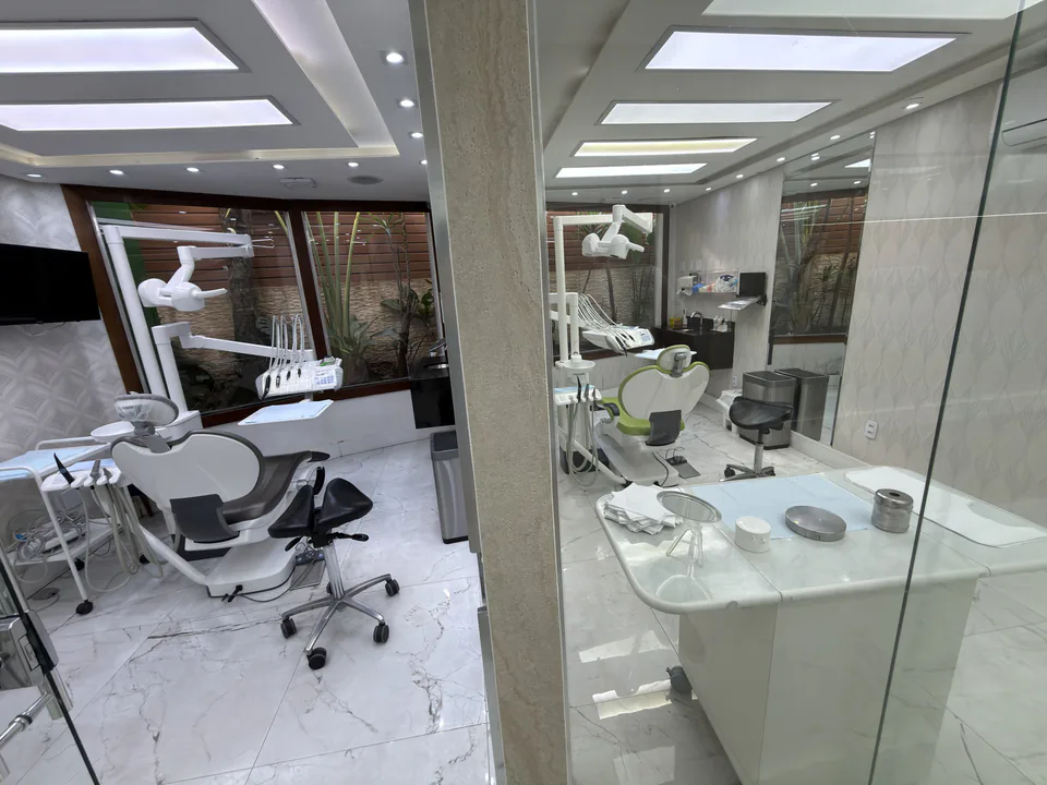
Two dental consulting rooms connected by a glass partition
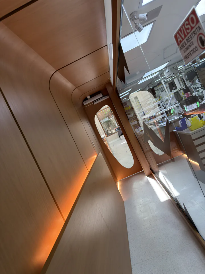
Curved corridor with wood panelling, amber lighting and a door at the far end

First, discover what has changed.

Then choose care that preserves what can be preserved and treats what genuinely needs intervention.

Talk to the Institute

Bibliographic references

Supporting sources for concepts, limitations and guidance. They do not replace individual assessment.

Original Portuguese content reviewed by Dr Silvana Puglisi, dental surgeon, CRO 33.773, technical director, on .

  1. Dhar V et al. Evidence-based clinical practice guideline on restorative treatments for caries lesions. Journal of the American Dental Association. 2023. PMID 37380250. DOI 10.1016/j.adaj.2023.04.011.
  2. Slayton RL et al. Evidence-based clinical practice guideline on nonrestorative treatments for carious lesions. Journal of the American Dental Association. 2018. PMID 30261951. DOI 10.1016/j.adaj.2018.07.002.
  3. American Association of Endodontists. Cracked Teeth. Patient guidance material, consulted in 2026.
  4. Watts A, Addy M. Tooth discolouration and staining: a review of the literature. British Dental Journal. 2001. DOI 10.1038/sj.bdj.4800959.
  5. National Institute of Dental and Craniofacial Research. Tooth Decay. National Institutes of Health. Official source.

Further reading

  1. ICDAS: International Caries Detection and Assessment System. ICDAS Coordinating Committee. ICDAS (2005). Official source

Privacy preferences

Your choice is valid for 180 days in this browser. You can change it under “Privacy”.

Forms may use Google reCAPTCHA to prevent abuse. Read the Privacy Policy