↗ PT

Other pages are available in Portuguese

Skip to content

Digital panoramic X-ray
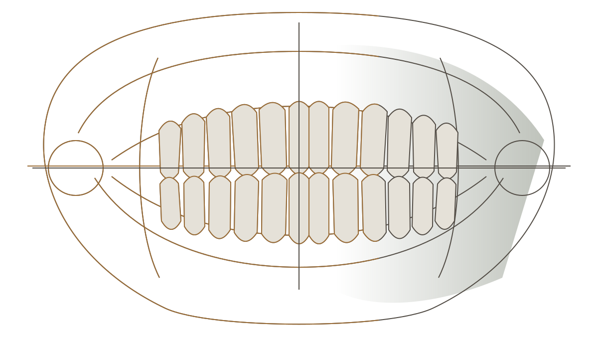
Conceptual representation of a panoramic dental field; it is not an actual radiograph or clinical case

A digital panoramic X-ray brings together teeth, the upper and lower jaws and nearby structures in a two-dimensional image. Its usefulness begins with a clear clinical question.

Discuss the examination

At Instituto Rafa Puglisi, the examination forms part of assessment. It does not replace the clinical consultation.
What a panoramic image shows

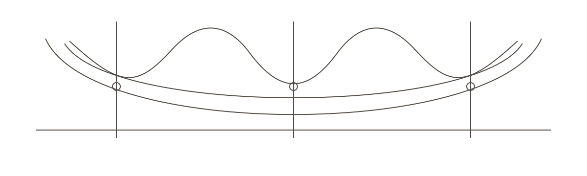
Three assessment points in a conceptual panorama: structures, relationships and context

The panorama brings an extensive area together in a single image. The clinical question guides interpretation and shows when another modality may be needed.

  1. Structures

    Teeth, the upper and lower jaws and nearby areas share the same field.

  2. Relationships

    The broad view helps locate regions and compare what is already present.

  3. Context

    History, clinical examination and the question guide interpretation. [1]

The indication comes before the image

The decision begins with history, clinical examination and the missing information. Radiography is used when its expected contribution justifies the exposure. [1]

Previous examinations are also part of that choice. Comparing existing images helps avoid unnecessary repetition.

Seeing a larger area does not mean reaching conclusions without context.

When another image may be needed

Panoramic radiography provides a general two-dimensional view. Another imaging examination may be recommended when a region needs more detail or the clinical question requires three-dimensional information.

How the examination is performed

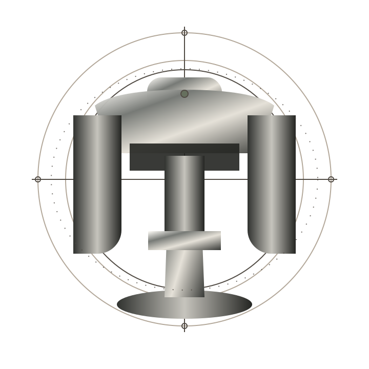
Conceptual representation of panoramic equipment. The model used may vary

Technology in the service of care

A useful image depends on examination selection, positioning and the question that prompted acquisition. The result is interpreted alongside the clinical information.

Brief
Acquisition takes just a few moments.
Positioned
Stability reduces avoidable distortion.
Interpreted
The image forms part of a broader assessment.

The examination is brief. Obtaining a useful image depends on preparation, alignment and remaining still during acquisition.

  1. Prepare

    The team advises removal of removable objects that interfere with the field, such as glasses, earrings and dentures when necessary.

  2. Position

    The head is supported and aligned in the equipment. Correct posture reduces avoidable distortion and overlap.

  3. Remain still

    The equipment travels around the head while the person holds their position for a few moments.

  4. Interpret

    The image is analysed alongside the clinical information that prompted the examination.

The lowest exposure needed for a useful image

The expected benefit must justify the examination. Technique, equipment and positioning need to produce diagnostic information and reduce repeat examinations. [2][3]

Tell the team about confirmed or possible pregnancy, recent examinations and any difficulty remaining still.

Questions before the examination

Does a panoramic image replace all other radiographs?

No. It provides a general view. Another imaging examination may be recommended when a region needs more detail or the clinical question requires three-dimensional information.

Do I need a panoramic X-ray at every appointment?

No. Need and frequency depend on individual assessment, history and the information that affects care. [1]

Does the examination hurt?

No sensor is placed inside the mouth. The person rests their face against the equipment supports and remains still during acquisition.

Do I need to remove earrings and glasses?

Removable metal objects near the area being examined interfere with the image. The team explains what needs to be removed before positioning.

What if pregnancy is confirmed or possible?

Tell the team before the examination. The professional assesses suitability, urgency, timing and protective measures according to the case.

What is the difference between a panoramic X-ray and a regular dental X-ray?

The panoramic X-ray shows the teeth, upper jaw and lower jaw in a single image, with nothing inside the mouth. X-rays taken with the film or sensor inside the mouth show a few teeth at a time, in more detail. That is why, for decay between the teeth, for example, an X-ray taken inside the mouth is usually more suitable. [1]

How long is a panoramic X-ray valid for?

There is no fixed expiry. A recent panoramic X-ray may be enough if nothing has changed since the exam. When there are new symptoms, treatments done or changes in the mouth, a new image may be needed. Bring your previous exams to the appointment. [1]

How much does a panoramic X-ray cost?

The Institute's panoramic X-ray is done for patients in treatment here, as part of diagnosis and planning. That is why there is no standalone price: it is included in the plan presented after the assessment. Treatment can be paid in up to 21 credit card instalments, depending on the treatment plan and current conditions.

Beyond aesthetics, we are a multidisciplinary clinic.

Explore options

Care takes place here

Across more than 400 m², seven consulting rooms and an integrated in-house laboratory bring together different stages of care at the same address. Discover the Institute.

A history in dentistry since 1923 connects generations with care today. Explore the Institute's history.

Documentary portrait of Dr Silvana Puglisi

Dr Silvana Puglisi is the dentist responsible for technical oversight. Meet our professionals who continue to provide care and carry on the work of LAB Puglisi.

Spacious reception at the Institute with artwork by Kobra
Lounge with green armchairs, artwork and a garden visible through the glass doors
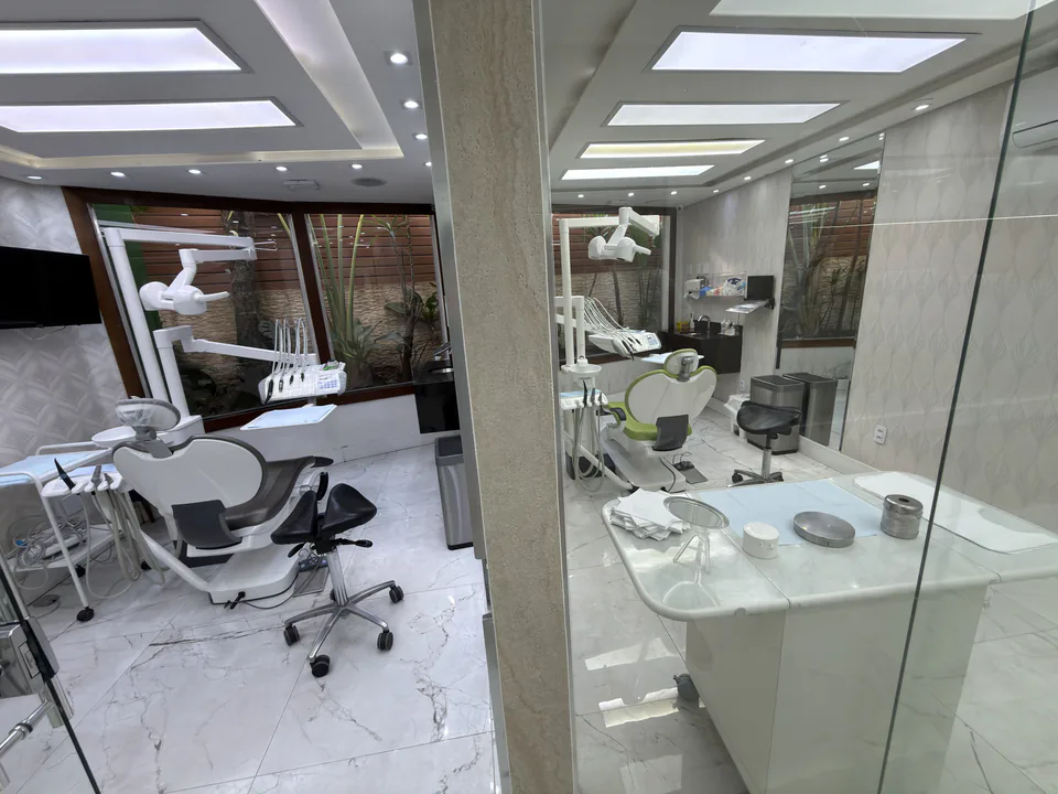
Two dental consulting rooms connected by a glass partition
Curved corridor with wood panelling, amber lighting and a door at the far end

The image gains meaning when it forms part of care

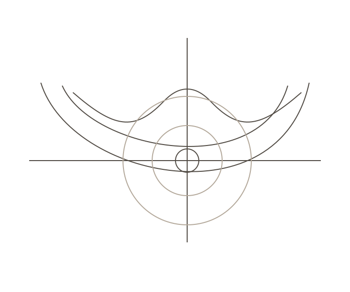
Conceptual arcs connecting the panorama to the clinical decision

If you want to understand which examination fits your assessment, talk to the Institute's team.

Discuss the examination

References and further reading

Original Portuguese content reviewed by Dr Silvana Puglisi, dental surgeon, CRO 33.773, technical director, on .

  1. American Dental Association and U.S. Food and Drug Administration. The Selection of Patients for Dental Radiographic Examinations. Recommendations revised in 2012.
  2. U.S. Food and Drug Administration. Medical X-ray Imaging. Principles of justification and exposure optimisation.
  3. International Atomic Energy Agency. Dental radiology: X-rays. Radiation protection in dentistry.

Further research

  1. Panoramic radiography in dentistry: current evidence. PubMed search. National Library of Medicine. Current results

General information for educational support. Suitability depends on individual clinical assessment.

Privacy preferences

Your choice is valid for 180 days in this browser. You can change it under “Privacy”.

Forms may use Google reCAPTCHA to prevent abuse. Read the Privacy Policy